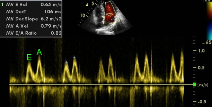
Normal E A Ratio Echo The E A ratio is a marker of the function of the left ventricle of the heart It represents the ratio of peak velocity blood flow from left ventricular relaxation in early diastole the E wave to peak velocity flow in late diastole caused by atrial contraction the A wave 1
Herein we present a comprehensive review of the echocardiographic early to late diastolic transmitral flow velocity E A ratio and the E to early diastolic mitral annular tissue velocity E e ratio placing each of these tests in clinical context for the practicing clinician An increased E e ratio has a direct significant relation with elevated left atrial pressure LAP in patients with depressed left ventricular systolic function However in patients with normal left ventricular systolic function E e does not appear to be useful 2
Normal E A Ratio Echo

Normal E A Ratio Echo
https://i.ytimg.com/vi/gM3GrF4E4jA/maxresdefault.jpg

Echocardiography Normal Vs Abnormal Images Heart Ultrasound Cardiac
https://i.ytimg.com/vi/yeYSX0Ps9I8/maxresdefault.jpg?sqp=-oaymwEmCIAKENAF8quKqQMa8AEB-AH-CYAC0AWKAgwIABABGGQgZChkMA8=&rs=AOn4CLAT92iE58OMDd3eV6TJIMMmID60LA

Diastole svg Cardiac Sonography Heart Echo Medical Photos Nursing
https://i.pinimg.com/originals/c0/2a/11/c02a11487ad80c946c6babd092007707.png
Which of following patients has the most advanced diastolic dysfunction or impaired relaxation Can he have a normal diastolic function LV diastolic parameters are altered in the presence of MAC This could be due to direct effects of MAC or might reflect truly reduced diastolic function Though there are several parameters for evaluation of left ventricular diastolic function by echocardiography the most commonly used are the pulsed Doppler mitral E A ratio and tissue Doppler mitral E e ratio
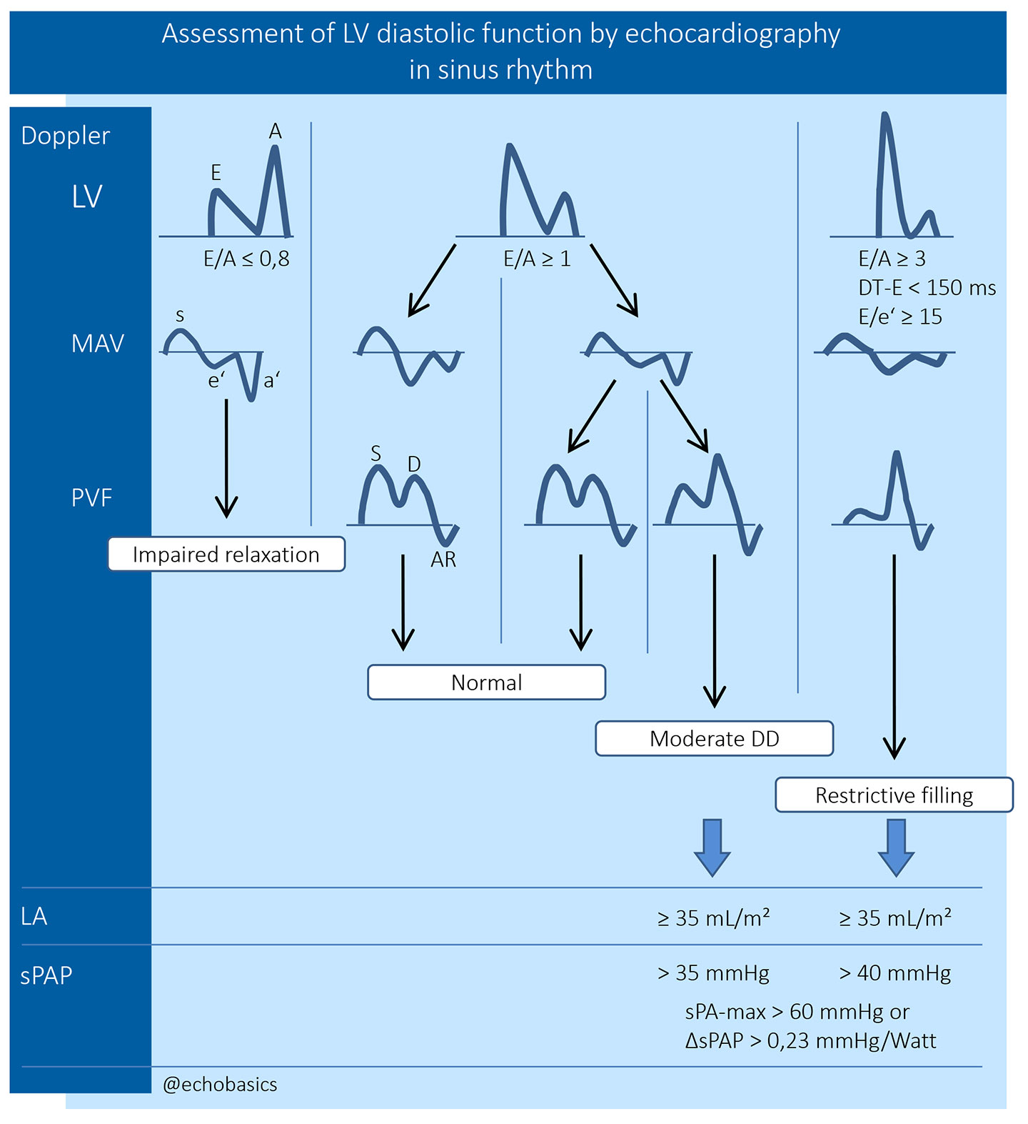
The E A ratio is a key measure when we evaluate for diastolic function The E wave represents early rapid filling and the A wave represents atrial contraction The ratio and pattern suggests if the diastolic filling is occurring at the proper time and rate during diastole To evaluate left ventricular diastolic function a PW Doppler sample volume is placed at the mitral valve leaflet tips and the following measurements recorded Three patterns or stages indicate abnormal diastolic filling The stage I filling pattern represents impaired slow
More picture related to Normal E A Ratio Echo

Rumus Cara Menghitung Berat Material Besi PDF 55 OFF
https://www.cardioserv.net/wp-content/uploads/2018/04/rap.png

EA Reversal All About Cardiovascular System And Disorders
https://johnsonfrancis.org/professional/wp-content/uploads/2015/01/EA-Reversal.jpg

Normal E A Ratio
http://www.fetalultrasound.com/online/text/7-092_files/image004.jpg
The presence or absence of diastolic dysfunction in patients with a normal LVEF is based on the assessment of four variables These variables and their cutoff values include If only 2 parameters are available if both are normal LAP is normal Consider the status of the left ventricle on two dimensional echo Even if the E A ratio and deceleration time are in the normal range LV filling is unlikely to be normal if there is significant LV hypertrophy or LV dysfunction In this setting preserved E velocity is better explained by elevated left atrial pressure than by brisk relaxation
[desc-10] [desc-11]

Pulsed Doppler In Fetal Echocardiography Obgyn Key Fetal Cardiac
https://i.pinimg.com/originals/f6/a8/40/f6a840ee1ce94ca2e7a43fe39a832687.jpg

Pin On Echocardiogram
https://i.pinimg.com/736x/51/2b/64/512b644a9930c4423ebe591d18e4f64b.jpg

https://en.wikipedia.org › wiki › E › A_ratio
The E A ratio is a marker of the function of the left ventricle of the heart It represents the ratio of peak velocity blood flow from left ventricular relaxation in early diastole the E wave to peak velocity flow in late diastole caused by atrial contraction the A wave 1

https://www.jacc.org › doi
Herein we present a comprehensive review of the echocardiographic early to late diastolic transmitral flow velocity E A ratio and the E to early diastolic mitral annular tissue velocity E e ratio placing each of these tests in clinical context for the practicing clinician

Mastering Diastology Part 1 Cardioserv

Pulsed Doppler In Fetal Echocardiography Obgyn Key Fetal Cardiac

Echobasics

Understanding The Basics LV Filling Patterns Cardioserv

Correct Techniques To Acquire Diastology Measurements

Mastering Diastology Part 2 Cardioserv

Mastering Diastology Part 2 Cardioserv

Mastering Diastology Part 2 Cardioserv

Finally Mitral Valve Orientation Explained Cardioserv

Normal Echocardiogram Results
Normal E A Ratio Echo - Which of following patients has the most advanced diastolic dysfunction or impaired relaxation Can he have a normal diastolic function LV diastolic parameters are altered in the presence of MAC This could be due to direct effects of MAC or might reflect truly reduced diastolic function