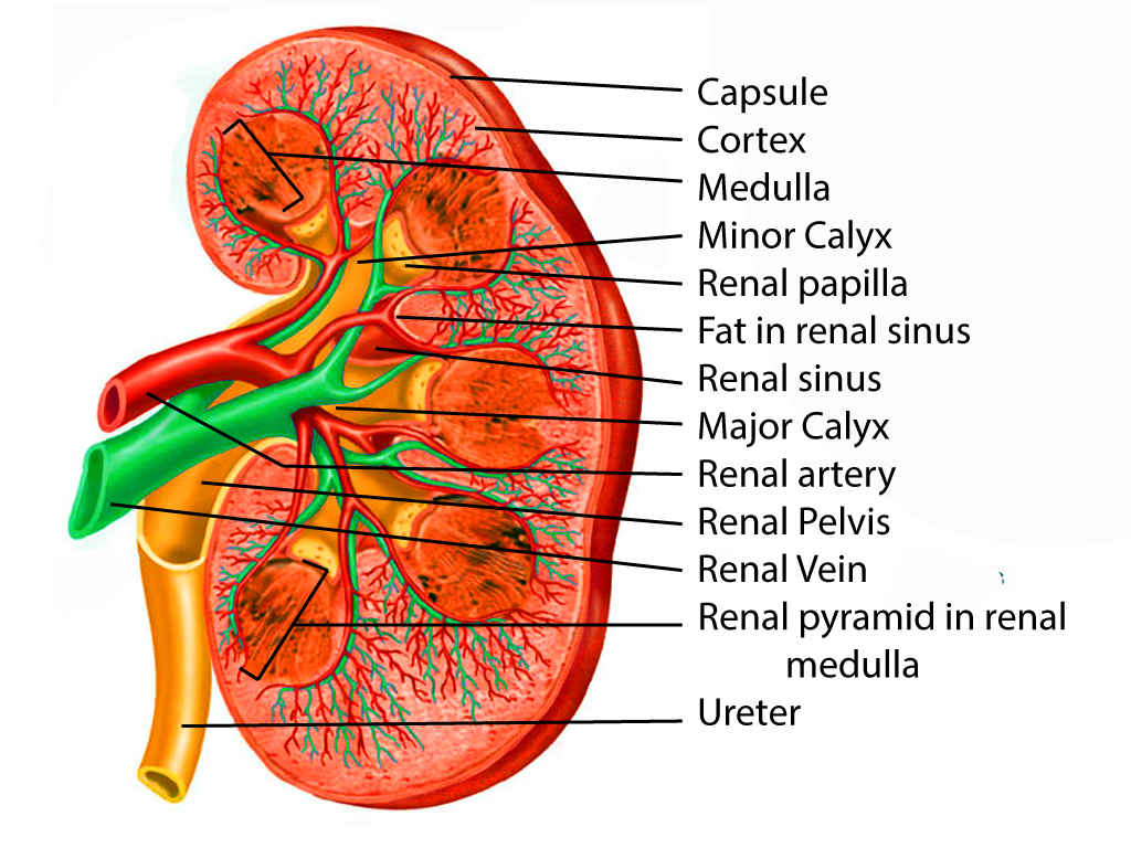
Renal Pelvis Size Each kidney normally has two or three major calyces which unite to form the renal pelvis The renal hilum is the entry to the renal sinus and lies vertically at the anteromedial aspect of the kidney
Kidney volume was calculated using the ellipsoid formula as Length x Width x Depth x 0 523 In this study the total renal volume was obtained by adding together both kidney volumes but without mentioning the separate values for the left and right kidney The renal pelvis is triangular in shape lies posteriorly in the renal hilum surrounded by fat and vessels and is formed by either the union of two to three major calyces or of seven to eleven minor calyces
Renal Pelvis Size
Renal Pelvis Size
https://f6publishing.blob.core.windows.net/aa5b2b52-5962-410a-b948-688733172771/WJN-8-44-g008.png

Abnormal Posiyion Of Renal Pelvis Interlobar Vein Renal Columns Renal
https://i.pinimg.com/originals/19/10/2d/19102d6fb19a5368e7d4ab70c5a9ca2b.png

How To Measure Kidney On Ultrasound Renal Length Width AP Thickness
https://i.ytimg.com/vi/JqJtnmPU8bM/maxresdefault.jpg?sqp=-oaymwEmCIAKENAF8quKqQMa8AEB-AH-CYAC0AWKAgwIABABGGUgZShlMA8=&rs=AOn4CLDaXMEkTkI-xYshVcyZrhcSp-GoJQ
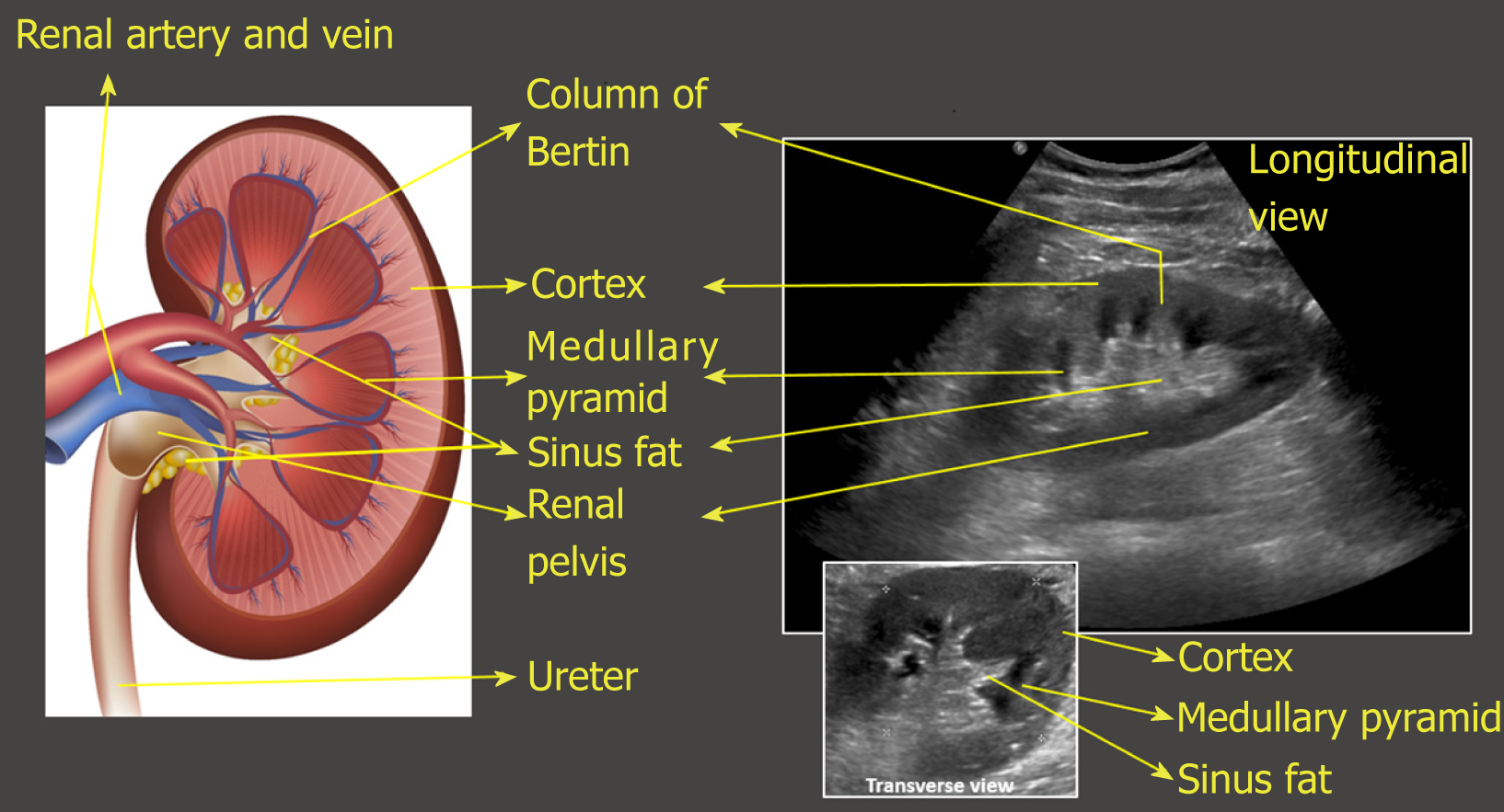
Reference charts of kidney length a kidney anteroposterior AP diameter b kidney transverse diameter c renal pelvis AP diameter d adrenal gland length e and kidney volume f showing raw data and fitted 5 th 50 th and 95 th centiles Kidney shape bean shaped with smooth contours parenchyma kidneys possess homogeneous echotexture with distinct corticomedullary differentiation renal pelvis central collecting system appears as hypoechoic structures with echogenic walls Pathological findings
The renal pelvis is the flattened superior end of the ureter It receives 2 or 3 major calyces each of which receives 2 or 3 minor calyces The minor calyces are indented by the renal papillae which are the apices of the renal pyramids A In adults the normal kidney is 10 14 cm long in males and 9 13 cm long in females 3 5 cm wide 3 cm in antero posterior thickness and weighs 150 250 g The left kidney is usually slightly larger than the right Diagram courtesy of Oregon State University
More picture related to Renal Pelvis Size

Kidney Size Chart For Renal Cyst
https://www.researchgate.net/publication/313259731/figure/tbl2/AS:614169678192661@1523440870880/Renal-length-of-both-kidneys-according-to-age.png

Measuring Anterior posterior Renal Pelvic Diameter APRPD In
https://www.researchgate.net/publication/357571834/figure/fig3/AS:1133720736149507@1647311495892/Measuring-anterior-posterior-renal-pelvic-diameter-APRPD-in-transverse-US-images-of-the.jpg

Renal Calculi Kidney Stones NCLEX Review
https://www.registerednursern.com/wp-content/uploads/2017/05/shutterstock_406914346-logika600-1024x1024.jpg
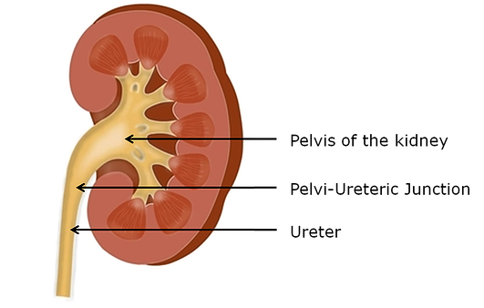
The renal pelvis is an expanded funnel shaped area through which urine travels It is located within the medial concave surface of the kidney filling the renal sinus The apex of the renal pelvis extends outwards from the kidney and becomes continuous with the superior end of Renal pelvis enlarged upper end of the ureter the tube through which urine flows from the kidney to the urinary bladder The pelvis is almost completely enclosed in the deep indentation on the concave side of the kidney the sinus
[desc-10] [desc-11]

Renal Ultrasound Made Easy Step By Step Guide Ultrasound Medical
https://i.pinimg.com/originals/4e/d8/91/4ed8913c0ac5e94c6fa518a6742f8336.png
Anatomy Abdomen And Pelvis Renal Artery Article StatPearls
https://www.statpearls.com/pictures/getimagecontent/61
https://radiopaedia.org › articles › kidneys
Each kidney normally has two or three major calyces which unite to form the renal pelvis The renal hilum is the entry to the renal sinus and lies vertically at the anteromedial aspect of the kidney

https://radiologyassistant.nl › pediatrics › normal...
Kidney volume was calculated using the ellipsoid formula as Length x Width x Depth x 0 523 In this study the total renal volume was obtained by adding together both kidney volumes but without mentioning the separate values for the left and right kidney

Feline Abdominal Ultrasonography What s Normal What s Abnormal Renal

Renal Ultrasound Made Easy Step By Step Guide Ultrasound Medical
Non dilated Renal Pelvis Measured In Antero posterior Dimension Slight

Ultrasound Screening Of The Kidneys And Urinary Tract In 11 887 Newborn

Change In Renal Pelvis Size In Obstructed Kidney Following IRE Of The

Renal Pelvis Anatomy Function Blockage Cancer Stone

Renal Pelvis Anatomy Function Blockage Cancer Stone
Level II Usg
Pelvi Ureteric Junction PUJ Obstruction Bristol Urology Associates

Normal Renal Vein Diameters Download Table
Renal Pelvis Size - Kidney shape bean shaped with smooth contours parenchyma kidneys possess homogeneous echotexture with distinct corticomedullary differentiation renal pelvis central collecting system appears as hypoechoic structures with echogenic walls Pathological findings
