Mildly Increased Bronchovascular Markings Noted At Bilateral Lung Fields Yahoo yahoo Yahoo Yahoo Yahoo
Yahoo yahoo Yahoo Mesno
Mildly Increased Bronchovascular Markings Noted At Bilateral Lung Fields

Mildly Increased Bronchovascular Markings Noted At Bilateral Lung Fields
https://i.ytimg.com/vi/dZ3wR0FrAXw/maxresdefault.jpg

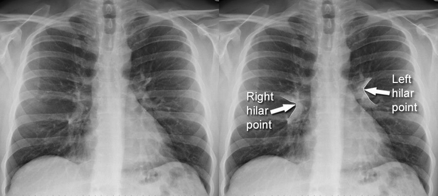
What Is Hilum How To View Hilar System In Xray Chest X 57 OFF
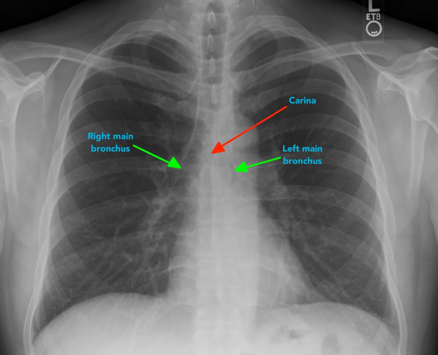
https://www.radiologymasterclass.co.uk/images/chest-images/cxr_aafde.jpg?mtime=20210304211312&focal=none

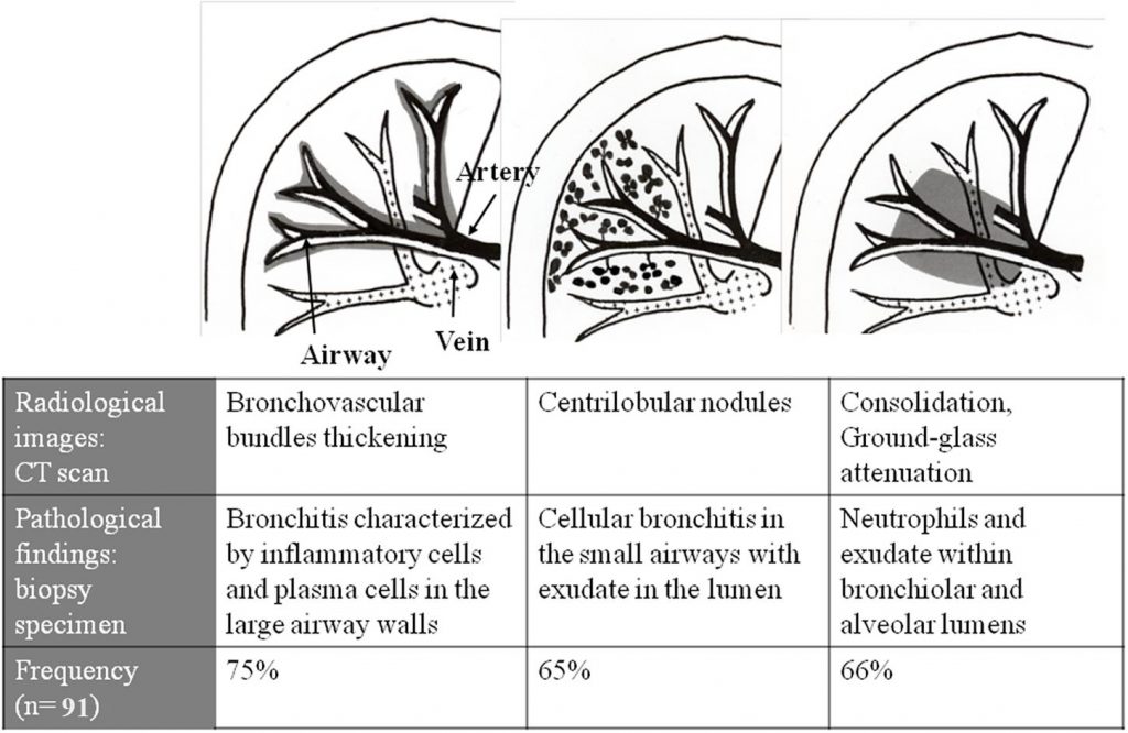
Sgowing Normal Bronchovascular
https://www.omicsonline.org/articles-images/CDPO-1-105-g001.gif
2021 11 1 Yahoo Yahoo 2011 1
yahoo cn yahoo cn Yahoo 2 28 2 28 2 28
More picture related to Mildly Increased Bronchovascular Markings Noted At Bilateral Lung Fields

Prominent Bronchovascular Structures Download Scientific Diagram
https://www.researchgate.net/profile/Oner-Ozdemir/publication/330614012/figure/fig2/AS:849500734251008@1579548166592/Prominent-bronchovascular-structures.png

Pulmonary Vascular Markings
https://www.researchgate.net/publication/316084219/figure/download/fig2/AS:625528499343360@1526149024368/Chest-radiograph-showing-increased-bronchovascular-markings-with-right-hilar-prominence.png

Pulmonary Vascular Congestion Cx Ray
https://www.researchgate.net/publication/343076471/figure/fig1/AS:915365265690629@1595251494102/Chest-X-ray-of-the-patient-The-image-displays-the-development-of-diffuse-pulmonary.png
Yahoo dev box 64 Linux 30 Yahoo 10 02 yahoo
[desc-10] [desc-11]

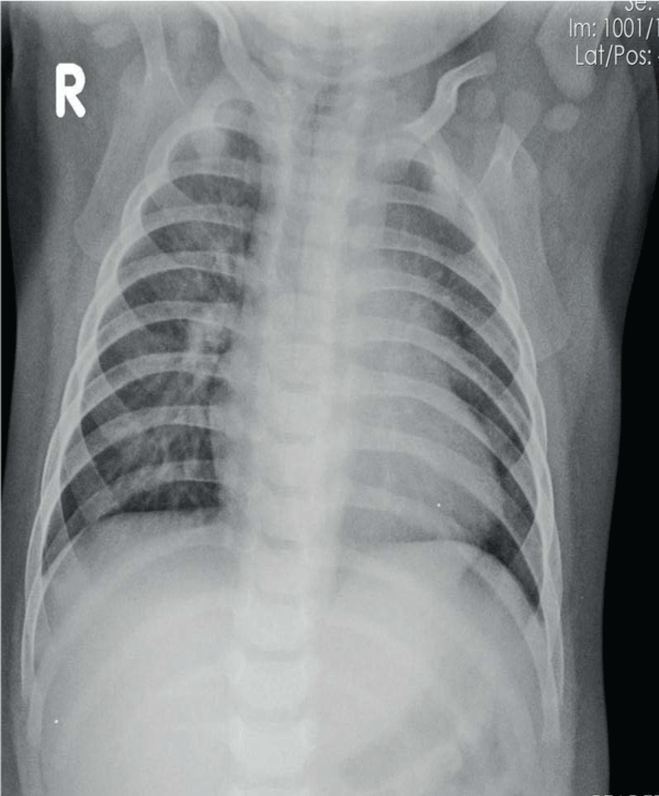
Peribronchial Cuffing Bronchiolitis Peribronchial Cuffing Reactive
https://i.pinimg.com/originals/9c/56/a1/9c56a1ad7df758f844e6414628b15bc1.jpg

Increase In Bronchovascular Markings With Multiple Inhomogeneous
https://www.researchgate.net/publication/371689259/figure/fig1/AS:11431281168993111@1687176943428/increase-in-bronchovascular-markings-with-multiple-inhomogeneous-opacity-in-both-lungs.png


X ray Chest Posteroanterior View A Showing Mildly Prominent

Peribronchial Cuffing Bronchiolitis Peribronchial Cuffing Reactive

Investigating Pleural Thickening The BMJ

What Is Peribronchovascular Distribution On CT Imaging Medicine

Chest Xray Showing Increased Bronchovascular Markings In Right

Raio X De T rax Interpreta o BRAINCP

Raio X De T rax Interpreta o BRAINCP
+-+Causes+and+Risk+Factors.jpg)
Interstitial Lung Disease Causes Symptoms And Treatment

Coronavirus Y Pulmones Humanos Sobre Fondo Blanco Vector Gratis

Diagnostic And Surgical Challenges In Disseminated Tuberculosis
Mildly Increased Bronchovascular Markings Noted At Bilateral Lung Fields - [desc-14]